


Essential Akasha Reset - 21-Day Cleanse Program
Anti-inflammatory doctor-protocol designed to help eliminate inflammation, restore your microbiome, achieve your optimal weight, balance hormones, and increase energy and vitality.
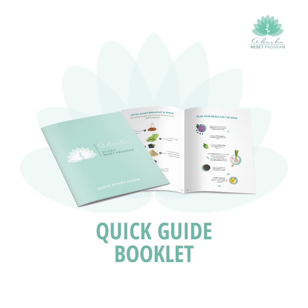
The Reset includes all the supplements and expert guidance you need, along with access to video lessons from our doctors and a handy printed guide packed with planning tools to keep you on course and detox safely.
“Inflammation is the common denominator in every disease. From a simple cold to cancer, inflammation is what links them.”

Low-grade inflammation can silently affect various aspects of our health, contributing to chronic conditions like heart disease, diabetes, and autoimmune disorders. This persistent inflammation can weaken the immune system, disrupt metabolic processes, and hormones over time.
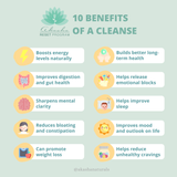
This program will reduce inflammation by eliminating toxins and inflammatory substances from the body, promoting the repair and regeneration of cells, enhancing immune function, and restoring balance to physiological processes.
The body runs its detoxification processes all day, every day, and the best way to help these happen efficiently is to provide the body with the nutrients essential to these critical functions.
21 days to embrace wellness with the Akasha Reset Program: reclaim your natural state of health and well-being. Following our eating protocol, natural supplements, and lifestyle changes, you will support your body's natural healing mechanisms.
The Anti-Inflammatory program developed by integrative doctors aims to restore Gut Health (Microbiome) and bring back your VITALITY.
Watch Dr. Maggie Ney explain why she recommends the Reset to her patients.
Supplements Included:
• UltraCleanse Plus - 33 oz
• Liver Detox - 60 Veggie Softgels
• Flora Plus Probiotics - 60 Capsules
• Digestazyme Digestive Support - 60 Tablets
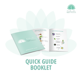
• Printed Quick Start Guide
• Exclusive Planning Tools
RETURNS POLICY
Return Policy: When buying Akasha Naturals products at the Akasha Center Clinic, you may return any unopened product within 30 days. Refunds will be credited back via the same method you used to pay. You may send the product along with the shipping receipt to the address listed on the outside of the package you received. You will be refunded as soon as our warehouse informs us of the returned package. It may take up to 10 days to see the amount in your account.


Doctor Formulated
From Our Board-Certified Medical Doctors & Practitioners

“Akasha Naturals is my go-to for supplements. I especially love the immune-support when I feel sickness coming on.”
INSTYLE Magazine

"Akasha helps cares for the health of my mind (my thoughts) and my spirit (my attitude about and in the life experience.”
Singer & Songwriter

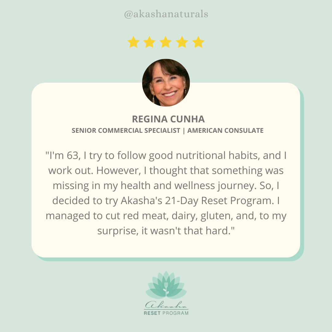
"By sticking to a strict diet and exercise plan, as well as natural remedies and Akasha supplements, I’ve lost over 40 pounds. I now have more energy and feel better than ever.”
Director & Screenwriter

"They are amazing and they work! I feel great and everyone comments about my level of energy and aliveness…Akasha Naturals are an investment in the life I love living."
Best-Selling Author

"With both the Akasha Center and Akasha Naturals, I’m happy I will never have to experience anything but strong and unwavering dedication to my care."
Writer & Director
Sustainably Sourced Supplements for a Healthier You and Healthier Planet
Our planet is our most precious resource. By choosing our supplements, you’re not only investing in your health and wellbeing, but also in the environment.We’re shaping a better future, through our commitment to people, the planet and purpose.